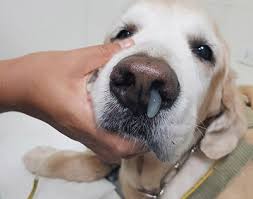
Canine distemper is a contagious, incurable, often fatal, multisystemic viral disease that affects the respiratory, gastrointestinal, and central nervous systems. Distemperis caused by the canine distemper virus (CDV).
Incidence
Canine distemper occurs worldwide, and once was the leading cause of death inunvaccinated puppies. Widespread vaccination programs have dramatically reduced its incidence.
CDV occurs among domestic dogs and many other carnivores, including raccoons, skunks, and foxes. CDV is fairly common in wildlife. The development of a vaccine inthe early 1960s led to a dramaticreductioninthe number of infecteddomestic dogs. It tends to occur now only as sporadic outbreaks.
Young puppies between 3 and 6 months old are most susceptible to infection and disease and are more likely to die than infected adults. Nonimmunized older dogsare also highly susceptible to infection and disease. Nonimmunized dogs that have contact with other nonimmunized dogsor with wild carnivores have a greater risk of developing canine distemper.
Transmission
Infected dogs shed the virus through bodily secretions and excretions, especially respiratory secretions. The primary mode of transmission is airborne viral particles that dogs breathe in. Dogsin recovery may continue to shed the virus for several weeks after symptoms disappear, but they no longer shed the virus once they are fully recovered.
It is possible for humans to contract an asymptomatic (subclinical) CDV infection. Anyone who has been immunized against measles (a related virus) is protected against CDV as well.
Signs and Symptoms
Macrophages (cells that ingest foreign disease-carrying organisms, like viruses and bacteria) carry inhaled CDV to nearby lymph nodes where it begins replicating (reproducing). It spreads rapidly through the lymphatic tissue and infects all the lymphoid organs within 2 to 5 days. By days six to nine, the virus spreads to the blood (viremia). It then spreads to the surface epithelium (lining) of the respiratory, gastrointestinal, urogenital, and central nervous systems, where it begins doing the damage that causes the symptoms of canine distemper.
Early symptoms include fever, loss of appetite, and mild eye inflammation that may only last a day or two. Symptoms become more serious and noticeable as the disease progresses.
The initial symptom is fever (103°F to 106°F), which usually peaks 3 to 6 days after infection. The fever often goes unnoticed and may peak again a few days later. Dogs may experience eye and nose discharge, depression, and loss of appetite (anorexia). After the fever, symptoms vary considerably, depending on the strain of the virus and the dog's immunity.
Many dogs experience gastrointestinal and respiratory symptoms, such as:
• Conjunctivitis (discharge from the eye)
• Diarrhea
• Fever (usually present but unnoticed)
• Pneumonia (cough, labored breathing)
• Rhinitis (runny nose)
• Vomiting
These symptoms are often exacerbated by secondary bacterial infections. Dogsalmost always develop encephalomyelitis (an inflammation of the brain and spinal cord), the symptoms of which are variable and progressive. Most dogsthat die from distemper, die from neurological complications such as the following:
• Ataxia (muscle incoordination)
• Depression
• Hyperesthesia (increased sensitivity to sensory stimuli, such as pain or touch)
• Myoclonus (muscle twitching or spasm), which can become disabling
• Paralysis
• Paresis (partial or incomplete paralysis)
• Progressive deterioration of mental abilities
• Progressive deterioration of motor skills
• Seizures that can affect any part of the body (One type of seizure that affects the head, and is unique to distemper, is sometimes referred to as a "chewing gum fit" because the dog appears to be chewing gum.)
Many dogs experience symptoms of the eye:
• Inflammation of the eye (either keratoconjunctivitis, inflammation of the cornea and conjunctiva, or chorioretinitis, inflammation of the choroid and retina)
• Lesions on the retina (the innermost layer of the eye)
• Optic neuritis (inflammation of the optic nerve which leads to blindness)
Two relatively minor conditions that often become chronic, even indogsthat recover are:
• Enamel hypoplasia (unenameled teeth that erode quickly in puppies whose permanent teeth haven't erupted yet—the virus kills all the cells that make teeth enamel)
• Hyperkeratosis (hardening of the foot pads and nose)
Inutero infection of fetuses is rare, but can happen. This can lead to spontaneous abortion, persistent infection in newborn puppies, or the birth of normal looking puppies that rapidly develop symptoms and die within 4 to 6 weeks.
Diagnosis
Diagnosis can be difficult and is based on the dog's vaccination history, clinical symptoms, and laboratory tests.
Blood tests usually are not helpful in the diagnosis, though insome cases they may reveal lymphopenia (a deficiency of lymphocytes, a type of immune system cell) during early infection, followed by leukocytosis (an increase in the number of white blood cells circulating through the blood) during later infection.
Imaging studies (e.g., x-rays, CT scans) can diagnose pneumonia.
Inclusion bodies (unique cellular structures that indicate the presence of the virus) can be detected with microscopic examination of buffy coat cells (cells that make up the "buffy layer" of centrifuged blood) and conjunctival secretions (secretions from the conjunctiva, the inner lining of the eyelids). A negative result does not rule out the possibility that the dog has distemper.
An immunofluorescent assay can detect viral antigens (proteins that the immune system manufactures to fight the virus) in the buffy coat cells and conjunctival secretions when inclusion bodies are not visible. Immunofluorescence involves using special proteins labeled with a fluorescent chemical that bind to the antigens and make them visible. Again, a negative result does not rule out the possibility that the dog has distemper.
Polymerase chain reaction (PCR), a technique that helps identify the virus's genetic material, is usually more sensitive than either microscopic examination for viral inclusions or immunofluorescence. It can be a difficult procedure and it is not always successful.
Cerebrospinal fluid (CSF) can be examined for CDV-specific antibodies and elevated levels of particular proteins and cells that indicate the presence of the virus.
Differential Diagnosis
Many diseases can cause symptoms resembling canine distemperand should be ruled out during diagnosis. Respiratory symptoms (e.g., cough and labored breathing) could be caused by bacterial pneumonia. Intestinal symptoms (e.g., vomiting and diarrhea) could be caused by gastroenteritis (an inflammatory bowel disease). Seizures and other neurological symptoms could be caused by toxoplasmosis (a protozoan infection) or epilepsy.
Treatment
Since there's no cure for distemper, treatment is supportive.
• Provide a clean, warm, draft-free environment.
• Keep eyes and nose clear of discharge.
• Give antiemetics (anti-nausea and anti-vomiting drugs) if there is vomiting.
• Give antidiarrheals for diarrhea.
• Monitor closely for dehydration. Dogswithout an appetite that are experiencing vomiting and diarrhea may require intravenous rehydration therapy.
• Antibiotics or bronchodilators are prescribed for pneumonia.
• Anticonvulsants may partially control seizures. Many veterinarians prescribe them before seizures start.
• Myoclonus is untreatable (and irreversible).
• Puppies who recover but have hypoplasia (unenameled teeth that erode quickly) can have the enamel restored to prevent further tooth decay.
• Glucocorticoid therapy can sometimes help blindness due to optic neuritis (inflammation of the optic nerve). This may help in the short term, but glucocorticoids weaken the immune system and may make symptoms worse.
Prevention
The best prevention against canine distemper is vaccination. Vaccination works well even in animals that have already been exposed to the virus, if it is administered within 4 days of exposure. Exposure to CDV via vaccination induces long lasting, but not permanent, immunity. Dogs should receive annual vaccinations to ensure protection.
Prognosis
Prognosis depends on the strain of canine distempervirus and the dog's immune response. After the initial fever subsides, the disease can progress ina number of ways.
The best prevention against canine distemper is vaccination. Vaccination works well even in animals that have already been exposed to the virus, if it is administered within 4 days of exposure. Exposure to CDV via vaccination induces long lasting, but not permanent, immunity. Dogs should receive annual vaccinations to ensure protection.
- There are several different types of distempervaccines available, each with advantages and disadvantages. Pet owners should discuss the various options with their veterinarians. The two most common vaccines are canine tissue culture-adapted vaccines and chick embryo-adapted vaccines.
- Canine tissue culture-adapted vaccines (e.g., Rockborn strain) are nearly 100% effective; they can very rarely cause fatal encephalitis (swelling of the brain) 1 to 2 weeks after vaccination. This type of vaccine is especially risky indogs with weakened immune systems.
- Chick embryo adapted-vaccines (e.g., Onderstepoort and Lederle strain) are safer than the Rockborn strain but are only about 80% effective.
- Most puppies are born with their mother's antibodies to CDV, which prevents them from becoming infected if exposed to the virus. They begin to lose their maternal protection between 6 and 12 weeks of age, which is when puppies should be vaccinated. Two to three vaccinations should be administered during this period. Dogs should be revaccinated yearly thereafter.
- Multidog households. Any dog that is suspected of being infected should be isolated from other dogs. Other dogs should be vaccinated, if they haven't already been.
- CDV doesn't last long outside the dog's body; heat, sunlight, most detergents, soaps, and various chemicals inactivate it. After an infected dog has been removed from the premises, contaminated objects and living areas should be disinfected with a 1:30 bleach-water solution.
Prognosis
Prognosis depends on the strain of canine distempervirus and the dog's immune response. After the initial fever subsides, the disease can progress ina number of ways.
- More than half of all dogs die between 2 weeks and 3 months after infection, usually from central nervous system complications. Most veterinarians recommend euthanasia for dogs that suffer progressive, severe neurological complications.
- Dogs that appear to recover may develop chronic or fatal central nervous system problems. Dogs with mild symptoms (e.g., myoclonus) may recover, though the symptoms can persist for several months or longer. Dogs with a strong immune response may never show any signs of infection. Once a dog has fully recovered, it no longer sheds the virus and is not contagious.


